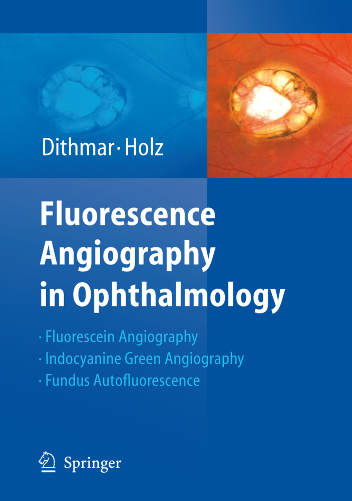
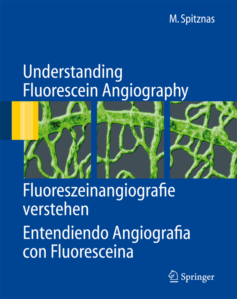
Fluorescence Angiography in Ophthalmology
Fluorescence Angiography in Ophthalmology
The technology of angiographic systems has been improved tremendously just within the past few years. This has allowed greatly increased levels of image resolution for both fluorescein and indocyanine green angiography.
This new atlas by Dithmar and Holz covers the basic principles of the new methods for fluorescein- and indocyanine green-angiography, as well as the high resolution imaging of fundus autofluorescence.
The angiographic signs of retinal and choroidal diseases are illustrated with images taken from a series of clinically relevant case examples that specifically illustrate the advantages of higher image resolution for the study of common retinochoroidal disorders. In so doing, this atlas offers an all-encompassing survey of the many angiographic signs in these disorders and their differential diagnoses. Clinicians in all subspecialties of ophthalmology can profit from a better understanding of these pathophysiological phenomena.
The physical and chemical fundamentals of fluorescence angiography
The Technical Fundamentals of Fluorescence AngiographyNormal Fluorescence Angiography and General Pathological Fluorescence Phenomena
Fundus Autofluorescence
Macular Disorders
Retinal Vascular Disease
Inflammatory Retinal/Choroidal Disease
Diseases of the Optic Nerve Head
Intraocular Tumors.
| ISBN | 978-3-662-51791-8 |
|---|---|
| Media type | Book |
| Edition number | Softcover reprint of the original 1st ed. 2008 |
| Copyright year | 2016 |
| Publisher | Springer, Berlin |
| Length | 224 pages |
| Language | English |

