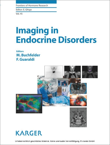
Imaging in Endocrine Disorders
Revolutionary changes in medical imaging have enormously improved the ability to detect structural and functional organ alterations early. Imaging is becoming an essential tool - in association with hormonal assays - for the diagnosis and management of endocrine disorders. New contrast media and their application to ultrasounds, as well as the opportunity to merge images acquired by functional/metabolic and traditional techniques, allow characterization of key features of identified lesions. Some radiological techniques such as ultrasonography, CT, and MRI are now available in operating rooms, thus supporting a diagnostic and therapeutic approach to endocrine diseases. In this new book, distinguished experts have contributed concise and well-illustrated chapters to describe pathognomonic features of several benign and malignant diseases affecting endocrine glands. They review the main advantages and disadvantages of each diagnostic technique along with indications for selecting a method. As a special feature, online videos of dynamic diagnostic and therapeutic procedures are available. Imaging in Endocrine Disorders is a must read and valuable reference for all professionals dealing with endocrine disorders, including internists and general practitioners who must manage the essential diagnostic workup.
1;Cover;1
1;Cover;1
2;Front matter;4
3;Contents;6
4;Preface;8
5;Sonography of Normal and Abnormal Thyroid and Parathyroid Glands;10
5.1;Abstract;10
5.2;Thyroid Ultrasonography;11
5.3;Parathyroid Ultrasonography;20
5.4;References;23
6;Computed Tomography and Magnetic Resonance Imaging of the Thyroid and Parathyroid Glands;25
6.1;Abstract;25
6.2;Thyroid;25
6.3;Parathyroid;28
6.4;References;31
7;Role of Nuclear Medicine in the Diagnosis of Benign Thyroid Diseases;33
7.1;Abstract;33
7.2;Nuclear Medicine: Radiopharmacology and Methods;34
7.3;Devices;34
7.4;Radiopharmaceuticals;35
7.5;Technetium-99m;35
7.6;Measurement of Technetium Uptake;35
7.7;Iodine Isotopes: 123 I and 131 I;35
7.8;Measurement of Iodine Uptake;36
7.9;Thyroid Scintigraphy;36
7.10;Other Thyroid Examinations and Tests;37
7.11;Benign Thyroid Diseases and Indications for Nuclear Medicine Tests;40
7.12;Acknowledgements;44
7.13;References;44
8;Hybrid Molecular Imaging in Differentiated Thyroid Carcinoma;46
8.1;Abstract;46
8.2;The Use of Radioiodine in Thyroid Carcinomas;47
8.3;Single-Photon Emission Computed Tomography/Computed Tomography in Differentiated Thyroid Carcinoma;49
8.4;Position Emission Tomography/Computed Tomography in Differentiated Thyroid Carcinoma;51
8.5;Conclusions;52
8.6;References;53
9;Endoscopic Ultrasound in Endocrinology: Imaging of the Adrenals and the Endocrine Pancreas;55
9.1;Abstract;55
9.2;Technical Details;56
9.3;Resolution;57
9.4;Indications and Typical Findings;57
9.5;Conclusion;62
9.6;References;62
10;Adrenal Imaging: Magnetic Resonance Imagingand Computed Tomography;64
10.1;Abstract;64
10.2;Introduction;64
10.3;Benign Lesions;70
10.4;Malignant Lesions;73
10.5;Image-Guided Intervention;75
10.6;References;77
11;Adrenal Molecular Imaging;79
11.1;Abstract;79
11.2;Molecular Imaging Techniques;80
11.3;Adrenal Incidentalomas;81
11.4;Molecular Imaging of Adrenal Tumors;82
11.5;Primary Aldosteronism;83
11.6;11 C-Metomidate PET in Adrenocortical Cancer;83
11.7;123 I-Metomidate SPECT;84
11.8;Pheochromocytoma;84
11.9;References;87
12;Gonadal Imaging in Endocrine Disorders;89
12.1;Abstract;89
12.2;Male Gonads;89
12.3;Female Gonads;97
12.4;References;104
13;Magnetic Resonance Imaging of Pituitary Tumors;106
13.1;Abstract;106
13.2;The Normal Pituitary Gland;106
13.3;Pituitary Adenomas;108
13.4;Collision Lesions of the Pituitary Region;120
13.5;The Postoperative Sella;120
13.6;Differential Diagnosis of Sellar Space-Occupying Lesions;123
13.7;References;128
14;Intraoperative Magnetic Resonance Imaging for Pituitary Adenomas;130
14.1;Abstract;130
14.2;Scanners and Setups;131
14.3;Results of Low-Field Studies;133
14.4;Results of High-Field Studies;135
14.5;Use of Neuronavigation;136
14.6;Intraoperative Imaging and Endoscopic Surgery;139
14.7;Conclusions;139
14.8;References;139
15;Molecular Imaging of Pituitary Pathology;142
15.1;Abstract;142
15.2;Receptor Imaging of Pituitary Tumors;143
15.3;Dopamine D2 Receptor Imaging of Pituitary Adenomas;143
15.4;Dopamine D2 Receptor Imaging of Pituitary Carcinomas;143
15.5;Somatostatin Receptor Scintigraphy of Pituitary Disorders;144
15.6;18F-Fluorodeoxy-D-Glucose Positron EmissionTomography and Pituitary Pathology;147
15.7;L - 11 C-Methionine Positron Emission Tomography and Pituitary Pathology;149
15.8;Conclusions;149
15.9;References;149
16;Imaging of Neuroendocrine Tumors;151
16.1;Abstract;151
16.2;Imaging Techniques;152
16.3;Neuroendocrine Tumor Characteristics, Presentationand Image Findings;156
16.4;Imaging in Therapy Monitoring;158
16.5;References;159
17;Author Index;161
18;Subject Index;162
19;Cover;166
Buchfelder
Guaraldi
Guaraldi
Ghigo
Benso
| ISBN | 9783318027389 |
|---|---|
| Artikelnummer | 9783318027389 |
| Medientyp | E-Book - ePUB |
| Copyrightjahr | 2016 |
| Verlag | Karger |
| Umfang | 156 Seiten |
| Sprache | Englisch |
| Kopierschutz | Adobe DRM |

