Ophthalmology for the Veterinary Practitioner
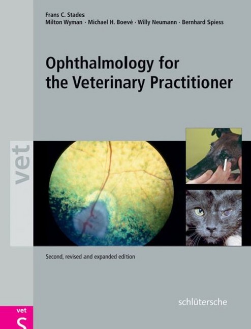
The standard work for ophthalmology in veterinary practise - now completely revised and extended. In its modern layout, the new edition continues a tried and tested concept. The present day developments in ophthalmological diagnostics and therapy are supplemented comprehensively throughout the book. In ophthalmology, the paramount maxim is 'Learning by doing'. This book provides a concrete guide to diagnostics and therapy of all important eye diseases in small animals especially for those general veterinary practitioners without any specific experience in ophthalmology and to students. It enables the determination of a diagnosis even without specialised instruments, while at the same time it gives answers to the important questions: When should an eye patient be referred to a specialist? Which preparations are necessary for doing this? Specific veterinary features in pet animals, horses, birds and farm animals are compared. A special chapter dedicated to ophthalmological emergencies enables quick and assured actions to be undertaken in emergency situations. The structure of the book follows the steps of a clinical examination making it particularly user-friendly. First-class new colour photographs and instructive drawings illustrate symptoms, diagnosis and treatment methods.
The authors All authors are well experienced in theory and practice since ages. They are one of the well respected Ophthalmologists in Europe and the USA. Frans C. Stades, DVM, PhD, Dip. ECVO Associate Professor of Veterinary Ophthalmology Department of Clinical Sciences of Companion Animals Faculty of Veterinary Medicine Utrecht University, The Netherlands Milton Wyman, DVM, MS, Dip. ACVO Professor of Veterinary Clinical Sciences Ohio State University College of Veterinary Medicine Professor of Ophthalmology Ohio State University College of Medicine Columbus, Ohio, USA Michael H. Boevé, DVM, PhD, Dip. ECVO Associate Professor of Veterinary Ophthalmology Department of Clinical Sciences of Companion Animals Faculty of Veterinary Medicine, Utrecht University, The Netherlands Honorary Professor of Veterinary Ophthalmology, Stiftung Tierärztliche Hochschule Hannover, Germany Willy Neumann, DVM, Dip. ECVO Specialist for Veterinary Surgery, Veterinary Ophthalmology Am Drosselschlag 25 Giessen-Heuchelheim, Germany Bernhard Spiess, DVM, PhD, Dip. ACVO/ECVO Department for Small Animals, Ophthalmology Unit Vetsuisse Faculty University of Zurich, Switzerland
1;Contents;6
2;Authors;11
3;Abbreviations;12
4;Origin of Plates and Figures;13
5;1 Introduction;14
6;2 Clinical and Differential Diagnostic Procedures;18
6.1;2.1 Description of the patient;18
6.2;2.2 Patient history;18
6.3;2.3 Animal handling, equipment, and instruments;21
6.3.1;2.3.1 Restraint and sedation;21
6.3.2;2.3.2 Materials and instruments;21
6.4;2.4 Examination of the eye and its adnexa;21
6.4.1;2.4.1 Head, skull, and orbital area;21
6.4.2;2.4.2 Tear film and tear production;22
6.4.3;2.4.3 Ocular discharge;23
6.4.4;2.4.4 Eyelids (palpebrae);23
6.4.5;2.4.5 Conjunctiva;24
6.4.6;2.4.6 Globe (bulbus);25
6.4.7;2.4.7 Sclera;26
6.4.8;2.4.8 Cornea;26
6.4.9;2.4.9 Anterior and posterior chambers;26
6.4.10;2.4.10 Pupil and iris;27
6.4.11;2.4.11 Lens;27
6.4.12;2.4.12 Vitreous;27
6.4.13;2.4.13 Fundus;27
6.4.14;2.4.14 Additional and specific examinations;28
6.5;2.5 Differential diagnosis;29
6.5.1;2.5.1 Introduction;29
6.5.2;2.5.2 The "red" eye;29
6.5.3;2.5.3 Epiphora without distinct blepharospasm;29
6.5.4;2.5.4 Blepharospastic / painful eye (Schirmer tear test not decreased);29
6.5.5;2.5.5 Protrusion of the nictitating membrane with enophthalmos;29
6.5.6;2.5.6 Exophthalmos;29
6.5.7;2.5.7 The "blue-white" cornea;30
6.5.8;2.5.8 The "pigmented" eye;30
6.5.9;2.5.9 The "blind" eye;30
7;3 Diagnostics and Therapeutics for Eye Diseases;32
7.1;3.1 Introduction;32
7.1.1;3.1.1 Into the conjunctival sac;32
7.1.2;3.1.2 Subconjunctival;34
7.1.3;3.1.3 Retrobulbar;34
7.1.4;3.1.4 Intraocular;34
7.1.5;3.1.5 General rules;35
7.2;3.2 Ocular therapeutic agents;35
7.2.1;3.2.1 Vasoconstrictors;35
7.2.2;3.2.2 Antihistamines (nowadays mostly replaced by corticosteroids);35
7.2.3;3.2.3 Antiglaucoma agents;36
7.2.4;3.2.4. Mydriatics;36
7.2.5;3.2.5 Antimicrobial agents;37
7.2.6;3.2.6 Corticosteroids;38
7.2.7;3.2.7 Non-steroidal anti-inflammatory drugs (NSAIDs);38
7.2.8;3.2.8 Local anesthetics;39
7.2.9;3.2.9 Vitamins, epithelializing agents,;39
7.2.10;3.2.10 Collyria;39
7.2.11;3.2.11 Other "drugs" for ocular use;39
7.2.12;3.2.12 Radiation;40
7.2.13;3.2.13 Protective devices;40
7.3;3.3 Surgical possibilities;40
7.3.1;3.3.1 Anesthesia;40
7.3.2;3.3.2 Preparation of the operative field;41
7.3.3;3.3.3 Positioning on the operating table;41
7.3.4;3.3.4 Draping;41
7.3.5;3.3.5 Magnification equipment;41
7.3.6;3.3.6 Surgical equipment;41
7.3.7;3.3.7 Suture material;41
7.3.8;3.3.8 Hemostasis;42
7.3.9;3.3.9 Cryosurgery;42
7.3.10;3.3.10 Laser techniques;42
8;4 Ocular Emergencies;44
8.1;4.1 Introduction;44
8.2;4.2 Luxation and proptosis of the globe;44
8.3;4.3 Chemical burns;47
8.4;4.4 Blunt trauma;47
8.4.1;4.4.1 Orbital fractures;47
8.4.2;4.4.2 Contusion of the globe;48
8.5;4.5 Penetrating or perforating trauma;50
8.5.1;4.5.1 Lid lacerations and conjunctival sac wounds;50
8.5.2;4.5.2 Conjunctival lacerations;52
8.5.3;4.5.3 Corneal lacerations;53
9;5 Orbital and Periorbital Structures;60
9.1;5.1 Introduction;60
9.2;5.2 Congenital abnormalities;61
9.3;5.3 Trauma;61
9.4;5.4 Enophthalmos;61
9.4.1;5.4.1 Enophthalmos due to loss of support;61
9.4.2;5.4.2 Enophthalmos due to Horner's syndrome;62
9.5;5.5 Exophthalmos;62
9.5.1;5.5.1 Exophthalmos due to swelling of the temporal muscles;63
9.5.2;5.5.2 Exophthalmos due to retrobulbar processes;63
9.5.3;5.6 Enucleation of the globe including the conjunctiva;66
9.5.4;5.7 Evisceration of the globe;69
9.5.5;5.8 Enucleation of the globe;69
9.5.6;5.9 Exenteration of the orbit;69
9.5.7;5.10 Orbitotomy;69
10;6 Lacrimal Apparatus;72
10.1;6.1 Introduction;72
10.2;6.2 Keratoconjunctivitis sicca (KCS);74
10.3;6.3 (Sialo)dacryoadenitis;77
10.4;6.4 Tear stripe formation;78
10.4.1;6.4.1 Micropunctum or stenosis of the lacrimal punctum;78
10.4.2;6.4.2 Atresia and secondary closure of the punctum;79
10.5;6.5 Dacryocystitis;80
10.6;6.6 Lacerations;83
10.7;6.7 Cysts and neoplasia;83
11;7 Eyelids;86
11.1;7.1 Introduction;86
11.2;7.2 Ankyloblepharon;
1;Front Cover;1 2;Back Cover;2 3;Copyright;6 4;Table of Contents;7 5;Body;15 6;Index;265
| ISBN | 9783899930955 |
|---|---|
| Artikelnummer | 9783899930955 |
| Medientyp | E-Book - PDF |
| Auflage | 2. Aufl. |
| Copyrightjahr | 2010 |
| Verlag | Schlütersche |
| Umfang | 272 Seiten |
| Sprache | Deutsch |
| Kopierschutz | Digitales Wasserzeichen |

